A young footballer twists the knee, and afterwards it swells, catches, and sometimes locks. An MRI shows a defect in the cartilage that lines the joint. Parents often expect this to heal like a bruise or a muscle pull. Cartilage is different, and understanding why is the first step to treating it well. The good news is that we have several effective options. The honest part is that none of them is a quick fix, and patience is part of the treatment.
Why cartilage is special, and stubborn
The ends of the bones in the knee are covered by a smooth, glistening layer called articular cartilage. It lets the joint glide almost frictionlessly and cushions load. Its strength is also its weakness: it has almost no blood supply. Muscle and bone heal well because blood carries the repair machinery to the injury. Cartilage, starved of that blood supply, has very little ability to repair itself once the surface is broken. A genuine defect usually will not simply fill back in with normal cartilage.
This is why a cartilage injury in a young, active person is taken seriously. Left unstable, a defect can enlarge, shed fragments that catch and lock the knee, and over time contribute to wear in that area. The aim of treatment is to settle symptoms and, where appropriate, restore a durable surface before more damage accumulates.
How we work out what you have
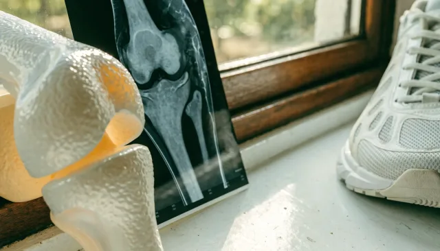
Diagnosis starts with the story (a twisting injury, swelling, catching or locking) and examination, and usually an MRI scan to map the size, depth and location of the defect and to check the rest of the knee, because cartilage injuries often travel with ligament or meniscus problems. Sometimes the full extent is only confirmed during a keyhole look inside the joint. You can read about keyhole knee surgery on the knee arthroscopy page.
There is no single best cartilage operation. The right choice depends on how big the defect is, where it sits, and who you are. Matching the technique to the lesion is the whole game.
The options, in plain terms
Treatment runs along a spectrum, chosen to fit the specific injury:
Non-surgical
Settle and rehabilitate
For small or stable lesions: activity modification, physiotherapy and time, while keeping a close eye on symptoms.
Arthroscopic tidy-up
Debridement
Keyhole removal of unstable flaps or loose fragments that catch and lock the knee, to settle mechanical symptoms.
Stimulate repair
Marrow stimulation
Techniques such as microfracture encourage the body to form a repair tissue over a contained defect.
Restore the surface
Transfer or cell repair
Osteochondral transfer (plugs of bone and cartilage) or cell-based cartilage repair for larger or deeper defects.
Just as important as the cartilage procedure is fixing what is around it. If the knee is unstable from an ACL tear, or a meniscus is torn, or the limb alignment overloads one side, those usually need addressing too, otherwise the new cartilage repair is set up to fail.
Realistic expectations
Cartilage repairs are not switch-on solutions. The repair tissue needs to mature, which means a protected, graded rehabilitation over months before full loading and sport. Outcomes vary with the size of the defect and how well the rest of the knee is managed. Many young athletes return to sport, but the ones who do best are the ones who respect the timeline rather than racing it.
What I tell young athletes and their families
Do not ignore a knee that swells, catches or locks, because a cartilage injury caught early keeps more options open and protects the joint. But also do not panic into the biggest operation. The best plan is the one tailored to your exact lesion and your goals, with the surrounding problems fixed at the same time, followed by a rehabilitation you commit to fully. Treated that way, a cartilage injury is a setback to manage, not the end of your sport.