On this page
Send the injury story, not only the scan
State when and how the injury happened. Describe the first swelling, whether you heard or felt a pop, and whether you could continue. Then explain what happens now: giving way, locking, pain, swelling after exercise and confidence on stairs or turns.
Add your sport, position, competition level, occupation and desired activities. A footballer who repeatedly pivots has a different decision from someone whose knee is stable during daily life.
For a settled injury, state whether the knee is improving with rehabilitation or still gives way. Repeated instability can damage the meniscus or cartilage, so the number and circumstances of later episodes matter.
What files are useful?
- The full MRI study in DICOM format
- The radiologist's written report
- Any X-rays and their reports
- A clinic or physiotherapy note, if available
- Your age, height, weight and injured side
- Previous injuries or operations
- Rehabilitation completed and the response
- Current medicines, allergies and relevant medical conditions
Why DICOM is better than a screenshot
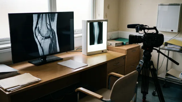
An MRI contains many sequences and slices. DICOM preserves the complete study and image quality. It allows the reviewer to move through the knee in order and inspect the ACL, menisci, cartilage, collateral ligaments and bone bruising.
A phone photograph often shows one selected slice with glare, compression or no identifying sequence. The report can identify a structural tear, but the original study lets the reviewer inspect that finding and the associated injuries. Both are useful for planning.
Does the age of the MRI matter?
An MRI taken soon after injury may show bone bruising and associated damage clearly. A later scan may be needed if the knee has sustained another giving-way episode, new locking or a fresh injury. The surgeon decides whether the existing study still answers the question.
Do not repeat an expensive scan automatically. Send the current study first, state what has changed and ask whether new imaging is useful.
What MRI can show
MRI can support the diagnosis of an ACL tear and show associated meniscus, cartilage, ligament and bone injury. It is highly useful but not perfect. A meta-analysis reported pooled sensitivity of about 87% and specificity of about 93%, with variation and study-quality limitations. [1]
Those figures describe test performance across groups. They do not mean a report is automatically correct or that imaging decides treatment.
Why X-rays may still be requested
MRI is strong for ligaments, menisci and cartilage, while X-rays show bone, alignment and established arthritis differently. A patient with a chronic ACL tear may have joint-space loss or other bony changes that affect the treatment discussion.
For younger patients, the surgeon may also need to know whether growth plates are open. Previous tunnels or fixation from an earlier reconstruction are often better understood with X-rays and, in selected revision cases, CT.
What cannot be tested through a screen?
Lachman and pivot-shift testing assess ligament behaviour. Examination also checks swelling, range, tenderness, strength, gait and other ligaments. Functional movement and confidence matter too. A video can show selected tasks, but it cannot safely reproduce the full hands-on assessment.
A torn ACL can look different over time
In the early weeks, pain and swelling may make the knee feel unstable even when the patient has not tested pivoting. Later, stronger muscles can make daily life comfortable while high-demand turns still cause giving way. A chronic injury may come with meniscal or cartilage damage that was not present on the first scan.
This is why the review asks both what the images show and what the knee currently does. Treatment is not selected from the word “complete” or “partial” in the report alone.
What the clinical history can add
The history helps clarify whether “instability” means true giving way, pain inhibition or fear after injury. The response to physiotherapy and the movements needed for sport or work also help explain the treatment options.
Prepare for the in-person visit
Continue the rehabilitation advised by your treating team, unless a locked knee or another urgent problem prevents it. Bring the original imaging, not just a web link that may expire. Write down the activities that cause giving way and the outcome you want from treatment.
Possible outcomes of the first review
- Continue structured rehabilitation and reassess stability.
- Obtain missing X-rays, better MRI files or another local examination.
- Discuss reconstruction because instability, activity and associated injury make it reasonable.
- Arrange urgent local care for a locked knee, major swelling or fracture concern; circulation or major new nerve symptoms need immediate emergency assessment.
- Travel for in-person assessment, with the final operation still subject to examination and clearance.
What the 24 to 48-hour response means
The preliminary remote review is free, with an initial response target of 24 to 48 calendar hours after usable records are complete. It can help identify the likely problem, any missing information and whether travel for assessment appears reasonable. Examination and medical clearance are still needed before an operation is confirmed.
If a link does not open or information is missing, the reviewing team should explain what is needed and help resolve the access problem. One organised record set makes the review easier.
Protect your medical information
The team can help you share the images and reports needed for review through the international contact route. Do not post identifiable medical information publicly.
Keep your own complete copy. You will need the same files for in-person assessment and for the physiotherapist or clinician who continues care at home.
An MRI tear is not an automatic operation
Some patients can return to their chosen activities without reconstruction after good rehabilitation. Others have recurrent giving way, want to return to pivoting sport or have associated repairable injury. The treatment decision brings the scan, examination, activity and goals together. [2]
For the broader decision, read ACL Tear: Do You Always Need Surgery? To submit a complete study, use the international patient form, email [email protected] or WhatsApp +91 80008 85008.
The records inventory can help separate files already available from those still needed. If something is missing, the team can help identify what is needed.
A final submission check
- Does the DICOM folder open?
- Is the written report included?
- Have you stated the injured side and injury date?
- Have you described giving way, locking and swelling?
- Have you listed your sport, work and goal?
- Have you included earlier injury, surgery and rehabilitation?
- Have you provided a safe way to contact you?