Most hip replacements are performed to restore movement and reduce pain. When a replaced hip becomes painful or unstable, however, there is no single explanation. The pattern and urgency of the symptoms guide the investigation.
On this page
Instability and dislocation
A dislocation occurs when the ball comes out of the socket. It may follow a fall or a particular movement, but some episodes have several contributing factors. Component position, soft-tissue tension, muscle function, spinal alignment, previous operations and patient factors can all matter.
A dislocated hip must be examined and imaged locally. Reduction is a medical procedure. Repeated instability later needs a structured assessment rather than repeated precautions alone. [7]
Loosening and bone loss
The cup, stem or both can lose stable fixation. Wear particles may lead to osteolysis, which is loss of bone around the components. Some patients notice gradually increasing groin or thigh pain. Others first present after a fracture through weakened bone.
Serial X-rays show whether the appearance is changing. CT may help define component position or bone loss. The scan is selected to answer the clinical question, not ordered as a routine bundle. [7]
Infection
Infection can occur early or years after hip replacement. Drainage, fever and worsening redness are concerning, yet slower infections may cause pain without these obvious signs. Blood tests and aspiration or culture may be needed. A normal-looking wound does not by itself exclude infection.
The distinction matters because infection surgery follows a different pathway. In some cases treatment is staged and takes much longer than a standard revision.
Fracture around the implant
A periprosthetic fracture often follows a fall, although weak bone and loosening can contribute. The fracture pattern, implant stability and available bone decide whether fixation, component revision or both are needed. New severe pain or inability to stand after a fall is an emergency. [7]
Wear and adverse tissue reactions
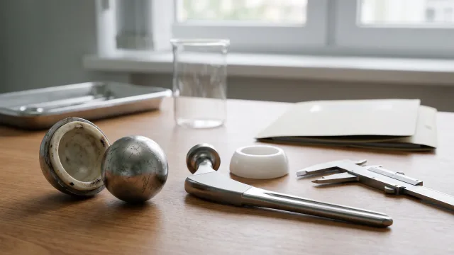
Every bearing produces some material over time. Modern bearings are designed to limit wear, but the amount and biological response vary. Wear can contribute to bone loss even before severe pain develops. [7]
Some component combinations raise additional questions about metal debris or local tissue reactions. This is a model-specific assessment. A blood metal-ion result or MRI finding should be interpreted with symptoms, implant identification and imaging rather than used alone to declare failure. [7]
Leg length and muscle problems
A feeling that one leg is longer can come from true length difference, pelvic tilt, spinal shape, muscle tightness or the way the patient walked before surgery. Measurement and imaging help separate these causes. Revision solely for a perceived difference should not be proposed without a careful assessment.
The abductor muscles help steady the pelvis during walking. Tendon damage or weakness may cause a limp, outer-hip pain or instability. Treatment may involve rehabilitation, tendon assessment or reconstruction, depending on the cause.
Why registry figures do not predict one person
Joint registries identify loosening, infection, instability and fracture among common reasons for hip revision. Their proportions vary by country, implant era, coding and length of follow-up. They describe groups. They do not tell an individual patient what has happened.
How a failed hip is assessed
- History: the original diagnosis, every operation, when the symptoms began and any falls, dislocations, wound problems or antibiotics.
- Examination: gait, leg length, strength, tenderness, movement and nerve and circulation status.
- Imaging: current and earlier X-rays, with CT or other imaging when needed.
- Infection work-up: blood tests and, where indicated, aspiration and cultures.
- Implant identification: operation notes, implant stickers, model, size and fixation.
Blood tests may screen for infection. Aspiration is considered when the history, examination, tests or imaging leave infection possible. Nuclear scans and metal-artifact-reduction MRI have selected roles, but they are not routine answers for every painful hip.
Pain near a hip replacement may come from somewhere else
Groin pain can arise from the joint, but tendons and the lower back can also refer pain there. Pain over the outer hip is often related to the abductor tendons or bursa. Thigh pain may be linked to the stem, the spine or another cause. Examination decides which direction deserves investigation.
This is one reason that a scan sent by email cannot confirm implant failure. Revision surgery will not solve pain that comes from the spine, a tendon or a medical condition outside the hip.
What is checked after a dislocation?
After urgent reduction, clinicians confirm that the joint is back in position and check for fracture or nerve problems. A planned instability review then considers how the episode happened, component orientation, soft-tissue tension, muscle function and the relationship between the spine and pelvis.
A first dislocation does not automatically lead to revision, and repeated dislocation does not have one standard operation. The treatment must address the reason the hip is unstable.
Why earlier imaging changes the picture
A comparison film may show that a component has moved gradually, that bone loss is progressing or that a fracture is new. It can also show that a finding was present while the hip felt well. Earlier films are useful alongside the latest one.
The original postoperative image is particularly helpful because it acts as a baseline. When only printed films are available, the reviewing team can help identify whether the radiology archive or patient portal can supply the original DICOM studies.
Not every abnormal image needs revision
An X-ray finding should be connected to symptoms and change over time. A stable component can have features that look unfamiliar to a patient but have not progressed. Conversely, early bone loss can deserve attention before it causes major pain.
Surveillance, rehabilitation or treatment of another pain source may be more appropriate than revision. When surgery is proposed, the plan should state which abnormality it will correct.
Questions to ask before planned revision
- Which diagnosis is the revision intended to correct?
- Has infection been investigated adequately?
- Which components are expected to remain?
- What bone loss or fracture reconstruction is anticipated?
- Could the plan become more extensive during surgery?
- What weight-bearing restrictions are possible?
- How will an urgent problem be managed after I return home?
What revision may involve
A stable component may sometimes be retained. In other cases the cup, stem or both must be removed. Bone loss can require graft, augments or specialised components. Selected instability revisions may use dual-mobility or constrained designs, but neither is a universal answer and neither creates zero dislocation risk.
Recovery can be very different from the first hip replacement. A limited liner or head procedure may permit early weight bearing, while major bone or fracture reconstruction may require protection. Infection treatment may involve more than one admission and has no standard travel timetable.
The records and tests checklist will help with a preliminary overseas review. If your problem is acute, seek local care before discussing travel. [2],[7]